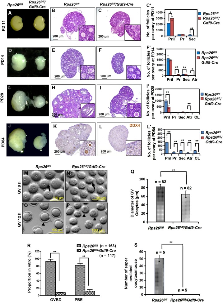
Fig. 2. Rps26 is essential for follicle development and oocyte growth.
a Ovaries of PD11 Rps26fl/fl/Gdf9-Cre mice and Rps26fl/fl mice were similar in size. b, c Hematoxylin/eosin staining of the paraffin slides of ovaries showing the similar morphologies of the ovaries at PD11 from Rps26fl/fl mice (b) and Rps26fl/fl/Gdf9-Cre mice (c). c’ Statistical analysis of the numbers of follicles in the ovaries of Rps26fl/fl mice (b) and Rps26fl/fl/Gdf9-Cre mice (c). *p < 0.05, **p < 0.01 as calculated by two-tailed Student’s t-tests. d Ovaries of Rps26fl/fl/Gdf9-Cre mice at PD14 were smaller than those of Rps26fl/fl mice. e, f The ovarian morphology at PD14 showing that there was arrest at the pre-antral follicle stage in the ovaries of Rps26fl/fl/Gdf9-Cre mice (f, insert) compared to Rps26fl/fl mice (e, insert). f’ Statistical analysis of the numbers of follicles in the ovaries of Rps26fl/fl mice (e) and Rps26fl/fl/Gdf9-Cre mice (f). *p < 0.05, **p < 0.01 as calculated by two-tailed Student’s t-tests. g, j Ovaries of Rps26fl/fl/Gdf9-Cre mice at PD28 (g) and PD84 (j) were smaller compared to ovaries of Rps26fl/fl mice. h, i Ovarian morphology in Rps26fl/fl/Gdf9-Cre mice at PD28 also showing arrest at the pre-antral follicle stage (i, insert) compared to the ovarian follicles in Rps26fl/fl mice that had developed into pre-ovulatory and antral follicles (h, insert). i’ Statistical analysis of the numbers of follicles in the ovaries of Rps26fl/fl mice (h) and Rps26fl/fl/Gdf9-Cre mice (i). **p < 0.01 as calculated by two-tailed Student’s t-tests. k, l Immunohistochemistry for the oocyte marker DEAD-Box Helicase 4 (DDX4) on ovarian slides of PD84 Rps26fl/fl/Gdf9-Cre mice (l, insert) showing no oocytes compared to those of Rps26fl/fl mice (k, insert). l’ Statistical analysis of the numbers of follicles in the ovaries of Rps26fl/fl mice (k) and Rps26fl/fl/Gdf9-Cre mice (l). **p < 0.01 as calculated by two-tailed Student’s t-tests. m, n GV oocytes collected from the ovaries of Rps26fl/fl mice (m) were larger in diameter than those from the ovaries of Rps26fl/fl/Gdf9-Cre mice (n) at PD21–28. o, p After 12 h in vitro culture, oocyte meiosis was arrested at the GV stage in Rps26fl/fl/Gdf9-Cre mice (p), while the oocytes of Rps26fl/fl mice had developed into MII oocytes (o). q The quantification of the diameters of GV oocytes shown in m and n showing that oocytes collected from the ovaries of Rps26fl/fl/Gdf9-Cre mice were significantly smaller than GV oocytes from the ovaries of Rps26fl/fl mice. **p < 0.01 as calculated by two-tailed Student’s t-tests. r Quantification of GVBD and PBE (shown in o and p) oocytes during meiosis. The proportions of GVBD and PBE were significantly decreased in vitro in the oocytes from the ovaries of Rps26fl/fl/Gdf9-Cre mice compared to those from Rps26fl/fl mice. **p < 0.01 as calculated by two-tailed Student’s t-tests. s Oocyte superovulation was inhibited in Rps26fl/fl/Gdf9-Cre mice compared to Rps26fl/fl mice. ** p < 0.01 as calculated by two-tailed Student’s t-tests

