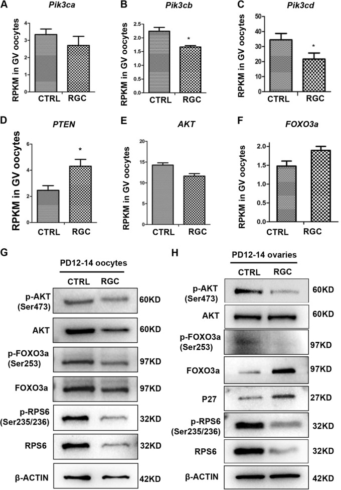
Fig. 6. The PI3K/Akt/Foxo3a signaling pathway was down-regulated in Rps26 knockout oocytes and ovaries.
a–c The mRNA expressions of Pi3k3ca (a), Pi3k3cb (b), and Pik3cd (c) were decreased in oocytes from PD14 Rps26fl/fl/Gdf9-Cre (RGC) mice compared to control (CTRL) Rps26fl/fl mice, as analyzed by RNA sequencing. *p < 0.05 as calculated by two-tailed Student’s t-tests. d, f The mRNA expressions of Pten (d) and Foxo3a (f) were increased in Rps26fl/fl/Gdf9-Cre (RGC) mice compared to control (CTRL) Rps26fl/fl mice. *p < 0.05 as calculated by two-tailed Student’s t-tests. e The mRNA expression of Akt was decreased in oocytes of Rps26fl/fl/Gdf9-Cre (RGC) mice compared to control (CTRL) Rps26fl/fl mice. g Western blot results showing the expression of p-Akt (Ser473), Akt, p-Foxo3a (Ser253), Foxo3a, p-Rps6 (Ser235/236), and Rps6 in the oocytes from Rps26fl/fl/Gdf9-Cre (RGC) mice and control (CTRL) Rps26fl/fl mice at the age of PD12–14. h Western blot results showing reduced expression of the p-Akt (Ser473), p-Foxo3a (Ser253), p-Rps6 (Ser235/236), and Rps6 proteins and increased expression of the Foxo3a and P27 proteins in the PD12–14 ovaries of Rps26fl/fl/Gdf9-Cre (RGC) mice compared to control (CTRL) Rps26fl/fl mice

