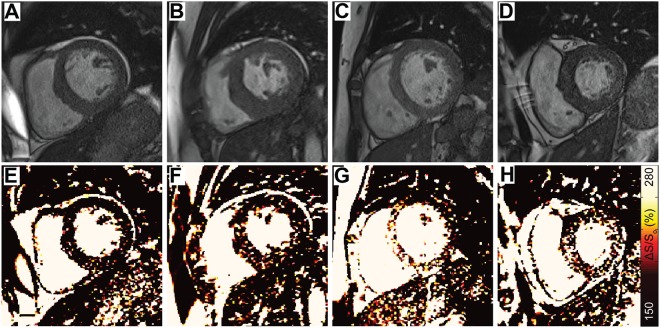
Figure 6.
Various spatial patterns of elevated ΔS/So were observed among hypertrophic patients with ESRD. (A–D) Representative end diastolic images acquired using a 45° excitation flip angle at the mid-ventricle are shown for 4 hypertrophic patients with ESRD. (E–H) Corresponding ΔS/So maps reveal different patterns and magnitudes of ΔS/So elevation despite the common structural feature of hypertrophy. An expanded version of this figure including 12 patients can be found in the Supplemental Data.

