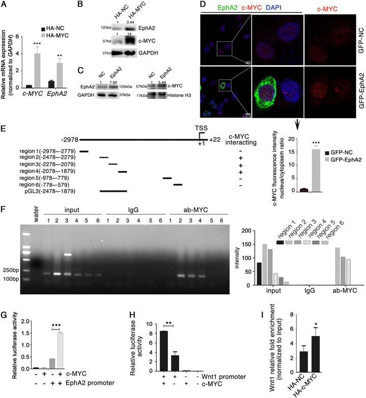
Fig. 4. EphA2 is a c-MYC target gene.
a, b c-MYC and EphA2 mRNA expression (a) and protein levels (b) in AGS cell lysates after transfection with the HA-tagged c-MYC expression vector. c Expression of nuclear and cytoplasmic c-MYC at 48 h post-transfection with Flag-EphA2 expression vector in AGS cells assayed by Western blotting. d Effect of EphA2 overexpression on the subcellular localization of c-MYC in AGS cells monitored by immunofluorescence. Cells were transfected with GFP-EphA2 or GFP-NC vector. e Schematic diagram of the truncated forms of the putative EphA2 promoter region and their interaction with c-MYC. f Identification of c-MYC-binding regions in the EphA2 promoter in AGS cells by ChIP using anti-c-MYC or normal rabbit IgG (negative control) (left panel). Quantification of band intensities of the PCR products is in right panel. g EphA2 transcription activity assayed in HEK293 cells transfected with the c-MYC expression vector and pGL3-EphA2-promotor luciferase reporter plasmid. h Wnt1 promoter-driven luciferase activity analyzed after co-transfection of HEK293 cells with the c-MYC expression vector and pGL3-Wnt1-promotor vector. i Identification of c-MYC-binding sites in the Wnt1 promoter in AGS cells by ChIP using anti-c-MYC. Normal rabbit IgG was used as a negative control. Relative accumulations of proteins in different groups compared with the negative control group are indicated. Significant differences were determined with the Student’s t-test. *P < 0.05, **P < 0.01, ***P < 0.001 compared with control group

