Fig. 5.
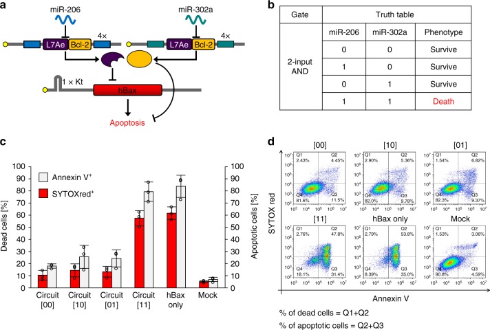
Apoptosis regulatory 2-input AND circuit. a The circuit has a pro-apoptotic gene, hBax, as the output. The anti-apoptotic gene Bcl-2 was fused with the L7Ae gene through P2A peptides to enhance the repression of apoptosis. b The truth table in the circuit is shown. For example, input pattern [10] means miR-206 present (=8 nM) and miR-302a absent (=0 nM). The circuit induces apoptosis (cell death) as output only when both miRNAs are present (=[11] state). c Cells were stained with SYTOX red for dead cell staining and Annexin V for apoptotic cell staining 24 h after the transfection. Data are represented as the mean ± s.d. (n = 3). Tukey’s multiple comparisons test results are shown in Supplementary Table 3. d Representative flow cytometry data of apoptosis regulatory AND circuits. The positive rate of SYTOX red was calculated by the sum of the percentage of the Q1 and Q2 fraction. The positive rate of Annexin V was calculated by the sum of the percentage of the Q2 and Q3 fraction

