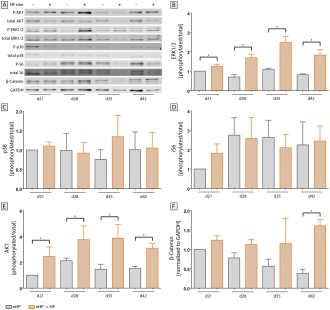
Figure 11.
Activation of crucial signaling pathways in osteoarthritis after hydrostatic pressure (HP) stimulation. (A) Representative immunoblots from the same gel were cropped to show specific bands for phosphorylated and total protein of Akt, ERK1/2, p38, ribosomal protein S6 as well as β-Catenin using GAPDH as housekeeping protein. Full-length blots are presented in Supplementary Figure S2. Pellets of unstimulated and HP-stimulated groups were harvested once every week for the last 21 days of the experiment and protein was isolated. Both (B) ERK1/2 and (E) Akt showed a statistically significant increase in activation in HP-stimulated pellets compared to static controls at all timepoints. (C) Protein expression of p38 did not significantly change over time. (D) Ribosomal protein S6, a downstream target of ERK1/2 and mTOR, did not exhibit an enhanced activation at any sampling timepoint. (F) Expression of β-Catenin increased towards the end of the stimulation period for stimulated pellets whilst it declined for unstimulated pellets. Mean + SEM; data from 2 individual donors, 6 replicates for each donor; *p < 0.05.

