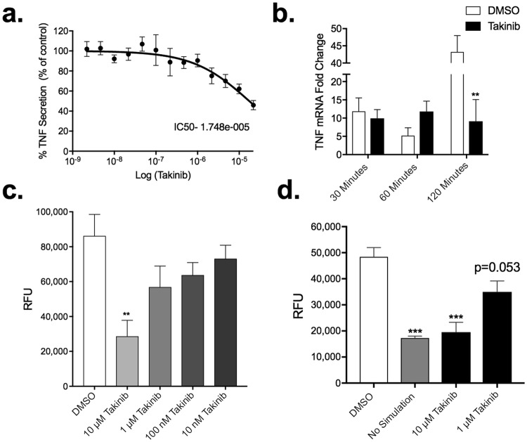
Figure 3.
Takinib reduces the functional response of macrophages upon pro-inflammatory activation with LPS and IFNγ. Raw264.7 cells were plated at (0.25 × 10⁶/well) in 24-well plates and activated with LPS (10 ng/mL) and IFNγ (50 ng/mL) and treated with the indicated Takinib concentration or vehicle control for 24 hours (a). RAW 264.7 cell were plated at (3 × 10⁶/plate) and serum starved for 4 hours. Cells were pretreated with either 10 μM Takinib or vehicle for 30 minutes followed by LPS (10 ng/mL) stimulation for the indicated times. mRNA levels compared to vehicle (n = 4± SEM) (b). Raw264.7 cells were plated at (0.75 × 10⁶/well) in a 96-well Boyden migration chamber in serum free media. Cells migrated towards, 10% fetal bovine serum, media chambers for 18 hours and total cell migration determined (c). (One way ANOVA with Dunnett’s, n = 3± SEM). Reduced NO production following LPS + IFNγ stimulation and treatment with Takinib in RAW 264.7 murine macrophage cells (One-way ANOVA with Dunnett’s, n = 3± SEM) (d). RFU = Relative Fluorescent Units *p < 0.05, **p < 0.01, ***p < 0.001.

