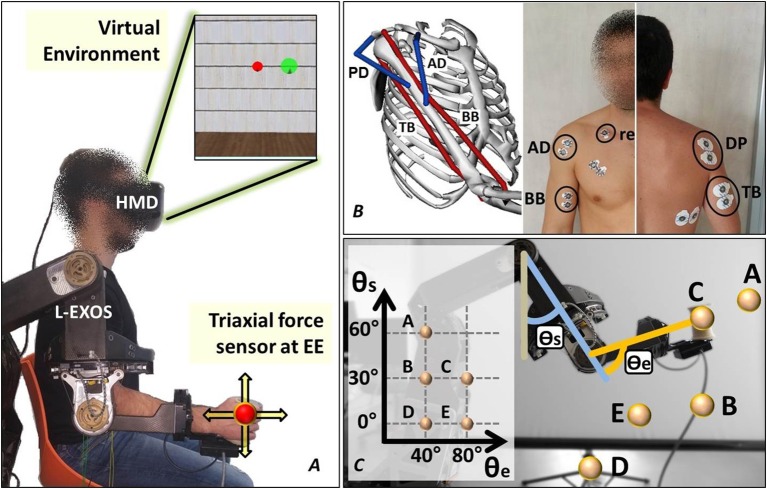
Figure 1.
The experimental setup. (A) The subject wears the upper limb exoskeleton controlled in position and the HMD displaying the cursor and the target spheres in a 3D virtual environment. (B) The neuromusculoskeletal model adopted in the study and the electrodes placement. (C) The five end-effector positions lying on the sagittal plane explored in the experiment.

