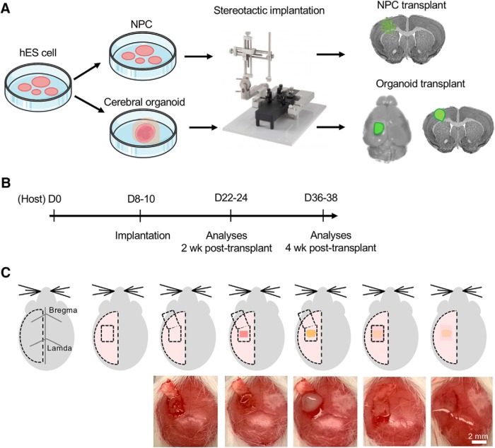
Figure 1.
Schematic depiction of intracranial transplantation of human cerebral organoids. A, hES cells were differentiated into NPC or cerebral organoids. Stereotactic surgery was performed to transplant one single cerebral organoid in the lesioned frontoparietal cortex in P8–P10 mice. For NPC transplantation, dissociated NPC were implanted into identical cortical region by stereotactic injection. B, Experimental timeline. P8–P10 immunocompetent mice were used as recipients for transplantation of dissociated human NPC or human cerebral organoids. Histologic analyses of the grafts were performed two or four weeks after grafting. C, Schematic diagrams (top) and intraoperative photographs (bottom) of cerebral organoid transplantation. Briefly, from left to right, once scalp is reflected, a small craniotomy window was opened with the bone flap hinged at the anterior base, and a small piece of the frontoparietal cortex (∼1 mm3) was removed. A single cerebral organoid was then implanted into the prelesioned mouse cortex, followed by return of the bone flap to close the craniotomy window, sealed with fibrin glue, followed by scalp closure.

