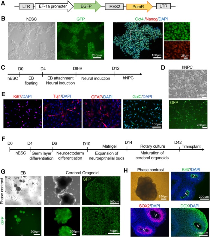
Figure 2.
Characterization of GFP-labeled NPC and cerebral organoids derived from hESC. A, Diagram of lentiviral vector expressing EGFP (enhanced GFP) driven by EF-1a promoter to label hES cells. B, Phase-contrast and fluorescent images of GFP-labeled hES cells (left) and expression of pluripotency markers Oct4 (green) and Nanog (red; right). C, Timeline of differentiation of hES cells into NPC. D, Phase-contrast and fluorescent images of human NPC derived from GFP-labeled hES cells. E, Representative immunofluorescence images of NPC stained for proliferation marker Ki67 and the indicated neural markers. F, Timeline of derivation of cerebral organoids from hESC. G, left panels, Phase-contrast and fluorescence images of GFP-labeled EB or Matrigel-embedded cerebral organoids. Right panel, Immunofluorescent images of sectioned cerebral organoids stained for GFP. H, Phase-contrast image of cerebral organoid at day 42 of culture and immunofluorescent images of sectioned day 42 cerebral organoid stained for the indicated markers. V: ventricle-like structures. Note the proliferative zone (Ki67+) and Sox2+ neuroprogenitors at the VZ/SVZ, and DCX+ neuroblasts in the outer layer.

