Figure 5.
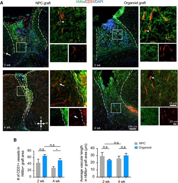
Vascularization of engrafted cerebral organoids. A, Representative immunofluorescence images demonstrate penetration of host CD31-positive blood vessels into hMito+ NPC grafts (left panels) or cerebral organoid grafts (right panels) at the indicated time points post-transplantation. Notice that CD31-positive endothelial cells inside the grafts are hMito-negative (white arrowheads). White arrows: host vasculature. Dashed white lines delineate the graft areas. Enlarged images of the boxed area are shown on the right. D: dorsal, V: ventral, M: medial, L: lateral. B, Quantifications of the number (left) and the average length (right) of CD31-positive blood vessels in hMito-labeled grafts demonstrate a higher number of vasculatures in the engrafted cerebral organoids compared to NPC transplants at four weeks after transplantation, but no significant difference in the average vascular length; *p < 0.05; n.s., non-statistically significant; n = 3 mice for each cohort, and at least two images analyzed from each mouse. Two-way ANOVA followed by a Tukey post hoc test.

