Figure 7.
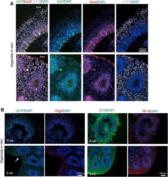
Stage-matched in vitro cerebral organoid characterization. A, Representative immunofluorescence images of cultured cerebral organoids at six or eight weeks of maturation show layered organization of cortical-like tissue with proliferating cells (Ki67+) and NPC (SOX2+) mainly localized in the VZ/SVZ and neurons (CTIP2+) localized in the outer layer. B, Representative immunofluorescence images of cerebral organoids after six or eight weeks of in vitro maturation. Left, A low number of astrocytes (GFAP+) was present in the organoids after eight weeks of maturation (white arrow), while no Olig2+ cells were detected. Right, Abundant neuroblasts (DCX+) were found at both time points while low level of expression of NF-H was detected at eight weeks of maturation.

