Fig. 1.

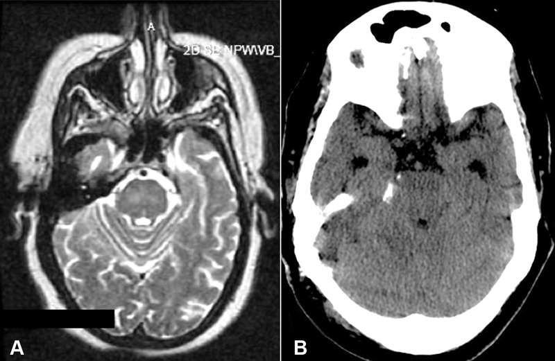
( A ) Preoperative T2-weighted axial magnetic resonance imaging (MRI); ( B ) Postoperative noncontrast axial computed tomography (CT) of brain.

( A ) Preoperative T2-weighted axial magnetic resonance imaging (MRI); ( B ) Postoperative noncontrast axial computed tomography (CT) of brain.