Fig. 6.
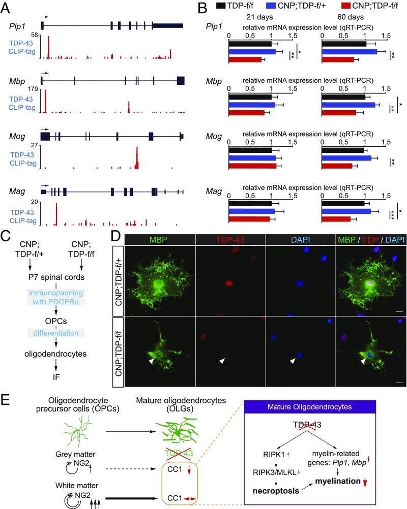
TDP-43 regulates the expression of myelin-related genes. (A) TDP-43 CLIP binding profiles for myelin genes. Peak heights represent overall read coverage. (B) Relative mRNA expression for myelin protein genes such as Plp1, Mbp, Mog, and Mag from the spinal cords of TDP-f/f, CNP;TDP-f/+, and CNP;TDP-f/f mice at both 21 d and 60 d of age as quantified by qRT-PCR. *P < 0.05, **P < 0.01, ***P < 0.001, one-way ANOVA. (C) Schematics for the immunopanning protocol to isolate and differentiate OPCs from P7 mice spinal cords. (D) Immunofluorescent staining of MBP (green) and TDP-43 (red) using differentiated oligodendrocytes derived from OPCs isolated from CNP;TDP-f/+ and CNP;TDP-f/f mice spinal cords, respectively. Oligodendrocytes without TDP-43 expression showed less complex MBP staining structure (arrowhead). (Scale bar: 10 μm.) (E) Graphic summary of a cell-autonomous role for TDP-43 in mature oligodendrocytes. TDP-43 depletion in mature oligodendrocytes leads to RIPK1-mediated cell-autonomous degeneration. Enhanced proliferation of OPCs compensates for the loss of mature oligodendrocyte in the white, but not the gray, matter. In parallel, TDP-43 regulates the expression of myelin-related genes, such as Plp1 and Mbp, that are essential for myelin. Deletion of TDP-43 down-regulates myelin-related genes, leading to loss of myelination capacity.

