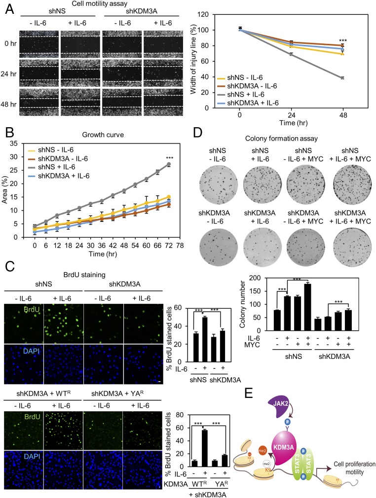
Fig. 4.
KDM3A phosphorylation is responsible for increased cell proliferation and motility. (A) (Left) Photomicrographs from the scratch-motility assay of HeLa cells expressing KDM3A shRNA with or without IL-6 treatment. Wound closure was monitored at 24-h intervals for 48 h in HeLa cells. (Right) Cell migration (percent) was quantified by calculating the wound width. ***P < 0.001 (Student’s t test). (B) Proliferation was monitored at 6-h intervals in HeLa cells ectopically expressing either control shRNA or KDM3A shRNA. Proliferation efficiency (percent) was quantified by calculating areas of cell population as shown in the graph. ***P < 0.001 (Student’s t test). (C) Confocal images of cells stained with BrdU. The fraction of increased BrdU-positive (BrdU+) cells after IL-6 treatment decreased following knockdown of KDM3A by shRNA. HeLa cells were knocked down by KDM3A shRNA and reconstituted with an shRNA-resistant form of KDM3A WT (WTR) or YA (YAR). Nuclei were counterstained with DAPI. (Scale bar, 10 μm.) ***P < 0.001 (Student’s t test). (D) (Top) Colony formation assay of HeLa cells transfected with either control shRNA or KDM3A shRNA in combination with MYC with or without IL-6 treatment. Cells were fixed and stained with crystal violet solution. (Bottom) Colony number was quantified as shown in the graph. ***P < 0.001 (Student’s t test). (E) Schematic model depicting JAK2−KDM3A−STAT3 signaling axis. Modulation of H3K9 methylation signature by JAK2-dependent phosphorylation of KDM3A is one of the predominant epigenetic events in transcriptional regulation of STAT3 target genes.

