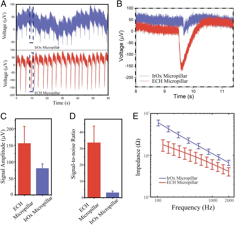
Fig. 3.
Electrophysiological recording with the ECH micropillar. (A) Extracellular recording of cardiomyocytes with conventional IrOx micropillar (Top) and soft ECH micropillar (Bottom) electrode with the same diameter of 3 μm. (B) Zoomed-in extracellular recording from the black box in A. (C) Signal amplitude and (D) signal-to-noise ratio of soft ECH versus IrOx micropillars. Error bars denote SD (IrOx n = 6; ECH n = 6). (E) Potentiostatic electrochemical impedance spectroscopy of ECH micropillars compared with IrOx micropillars with the same diameter at electrophysiologically relevant frequencies. Error bars denote SEM (n = 49 IrOx, n = 50 ECH).

