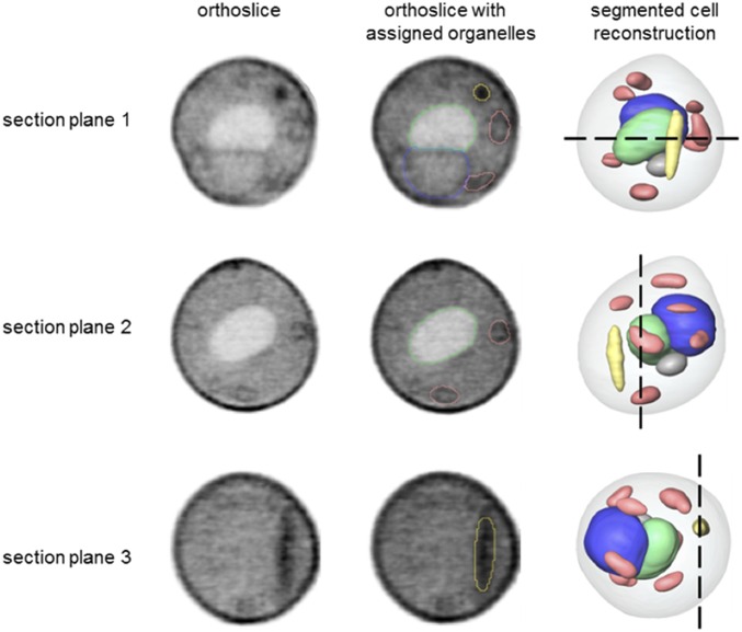
Fig. 5.
Ultrastructural features of S. cerevisiae–E. coli ΔnadA chimera cells. Segmented reconstruction of a S. cerevisiae–E. coli ΔnadA cell viewed from three different perspectives (section planes 1–3). Orthoslices (Left) at positions indicated by the black dashed lines in a reconstructed cell (Right) are shown for each perspective. The same orthoslices are overlaid in the Center column with outlines indicating segmented organelle assignment. The gray values were generated using LACs, with black corresponding to the highest LAC value. Organelle color key: green, vacuole; blue, nucleus; salmon, mitochondria; and yellow, high LAC value, bacteria-like structure that remained unassigned after segmentation.

