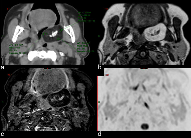
Figure 1.

CT scan (a) and MRI [(b) axial T1 weighted; (c) axial STIR; (d) axial diffusion-weighted imaging with background suppression) show a juxtatonsillar left parapharyngeal mass with central calcification and fat tissue peripherally. Hounsfield units in the CT scan ranged between –80 and –116 (a), while fat saturation by means of STIR technique (c) confirmed the presence of fatty tissue. CT scan after ontrast media showed no enhancement (not shown). STIR, short tau inversion-recovery.
