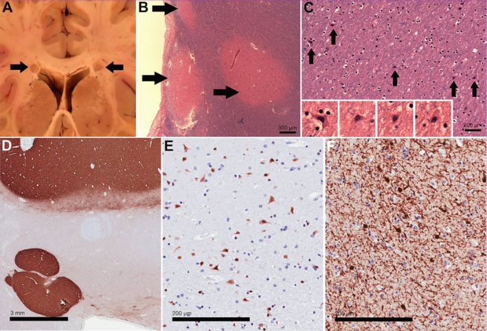
Figure 2.

Neuropathologic features in patients with 22q11.2DS (A–C from Patient 6, D–F from Patient 7). (A) Macroscopic image of coronal section through the brain showed bilateral PNH adjacent to the lateral ventricles in the white matter of both frontal lobes. Size of heterotopias was approximately 4 mm. (B) Histologic sections of the PNH (arrows) revealed disorganized aggregates of gray matter containing haphazardly arranged neurons. (C) Frequent individual heterotopic neurons were detected in the white matter surrounding the heterotopias. Magnification (insets) reveals cytologic details, such as pyramidal shapes. (D) Immunohistochemistry for synaptophysin in a section containing two heterotopic nodules underlines the fact that the nodules are composed of gray matter. (E) Immunohistochemistry for NeuN labels the neurons in the nodule, suggesting that they are fully differentiated. (F) Higher magnification of nodule with immunostain for Calretinin, a marker of cortical interneurons.
