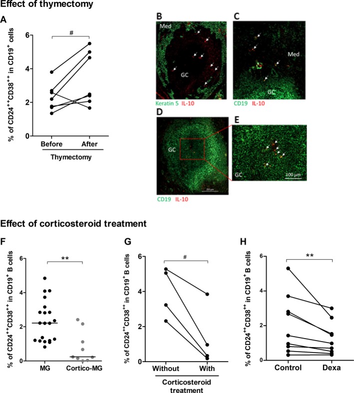
Figure 2.

Impact of classical MG therapies on the proportion of Breg cells. Effect of thymectomy: (A) PBMCs from seven MG patients were labeled before and 6–24 months after thymectomy to measure the percentage of CD24++ CD38++ in CD19+ B cells. P‐value was assessed by the paired t‐test (# P = 0.05). Only one patient had an inversed behavior with a decreased percentage of Breg cells after thymectomy. We had no reason to exclude the patient from the analysis but this patient also behave differentially regarding the % of regulatory T cells and Th17 cells compared to other patients. (B–E) Immunofluorescence staining of thymic sections from MG patients with an anti‐CD19 antibody for B cells or an anti‐keratin 5 for thymic medullary epithelial cells (in green), and anti‐IL10 (in red). Images were acquired with a Zeiss Axio Observer Z1 Inverted Microscope. Effect of corticosteroids: (F) PBMCs from MG patients (n = 20) and cortico‐treated MG patients (n = 9) were labeled to analyze the percentage of CD24++ CD38++ cells in the CD19+ B cells. P‐value was assessed by the Mann–Whitney test (**P = 0.002), (G) PBMCs from four MG patients followed up during in the course of their corticosteroid treatment were labeled to measure the percentage of CD24++ CD38++ in CD19+ B cells. P‐value was assessed by the paired t‐test (# P = 0.02). For three patients, the blood samples were initially without treatment and 1–24 months later while they were under corticosteroid treatment. For one patient, the blood sample was initially with corticosteroid treatment and 3 months later without treatment. (H) PBMCs from healthy controls were cultivated 48 hours with dexamethasone (Dexa) or without (control) and the percentages of CD24++ CD38++ cells in the CD19+ B cells were analyzed by flow cytometry. P‐value was assessed by the paired t‐test (**P = 0.008).
