Figure 2.

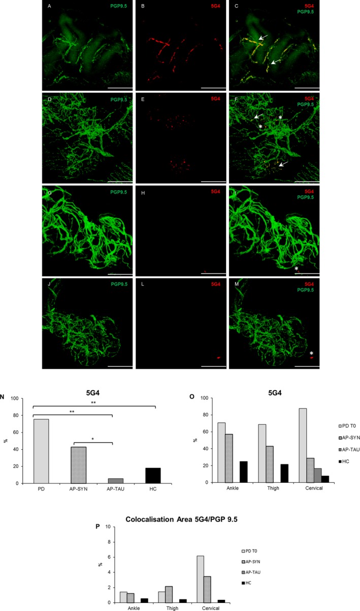
5G4 deposits are more expressed in PD and AP‐SYN patients. Confocal images of immunofluorescence with PGP9.5 (green) and 5G4 (red) of dermal nerves around sweat glands in PD T0 (A–B), AP‐SYN (D–E), AP‐TAU (G–H), and healthy subject (J–L). In yellow colocalization of 5G4 and PGP 9.5 (C,F,I,M) along axons. Scale bar 50 μm. White arrows indicate positive structures, asterisks indicate unspecific staining in non‐neuronal structures. The percentage of 5G4 is higher in PD T0 compared to HC and AP‐TAU (P FDR < 0.012), but not compared to AP‐SYN, and in AP‐SYN more than in AP‐TAU (P FDR = 0.04) (N); no significant differences among localizations but a tendency of higher 5G4 at cervical site in PD (O). A major colocalization area 5G4/PGP9.5 was measured in PD with a proximal to distal gradient (P).
