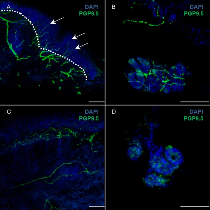
Figure 4.

Skin innervation: intraepidermal nerve fiber density (IENFD). Immunofluorescence staining with anti‐PGP9.5 (green) and DAPI (blue) of cervical skin in an healthy subject (A) and PD (C); in A the dotted white line shows the border between epidermis and dermis: IENFD is calculated as the number of nerve fibers (arrows) crossing the border per mm. Dermal nerve fibers around sweat glands are shown in an healthy subject (B) and PD (D). The pictures shows skin denervation in PD more evident at level of autonomic nerve fibers of sweat gland. Scale bar 50 μm.
