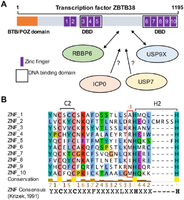
Figure 1.

Domain structure of ZBTB38. (A) The N-terminal BTB domain, the 10 zinc fingers, and the domain of interaction with RBBP6 and USP9X are schematized. DNA binding domains are also highlighted. Under the scheme, E3 ligases and deubiquitinases that may contribute to the regulation of ZBTB38 stability are listed. (B) Multiple sequence alignment of ZBTB38 zinc fingers using online service MAFFT and visualized using Jalview.17 A conserved consensus amino acid sequence is given at the bottom of the alignment. The position of the conserved lysine/arginine amino acids in DNA binding ZNFs is indicated (red rectangle). Conserved amino acids are colored according to residue type.
