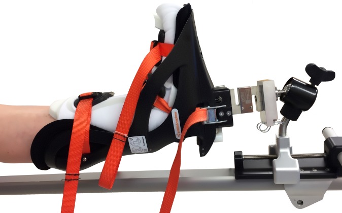
Figure 1.
Image of the custom attachment that was created to allow integration of an S-type load cell into the traction system. The patient was positioned on the surgical table and traction system. After anesthesia induction, a fluoroscopic image was obtained to define the pretraction joint position. Traction was then applied to the surgical limb, and a second fluoroscopic image was acquired in the same plane to confirm that sufficient distraction had been achieved to allow completion of all components of hip arthroscopic surgery in the central compartment. Traction force was continuously measured through this process.

