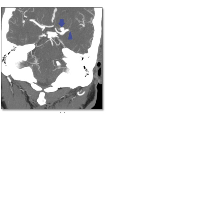
Figure 2.
A 66-year-old man with a left M1 occlusion and an incidental 7 mm middle cerebral artery aneurysm just proximal to the site of occlusion. The first image is a maximum intensity projection from the computed tomography angiogram showing a 7 mm aneurysm (arrow) adjacent to the site of occlusion (arrowhead). Intraprocedural angiogram (second image) during stroke intervention shows the aneurysm (arrow) and site of occlusion (arrowhead). The third image is a control angiogram showing successful recanalization of the previously occluded middle cerebral artery after mechanical thrombectomy (arrowhead) as well as the left middle cerebral artery aneurysm (arrow).

