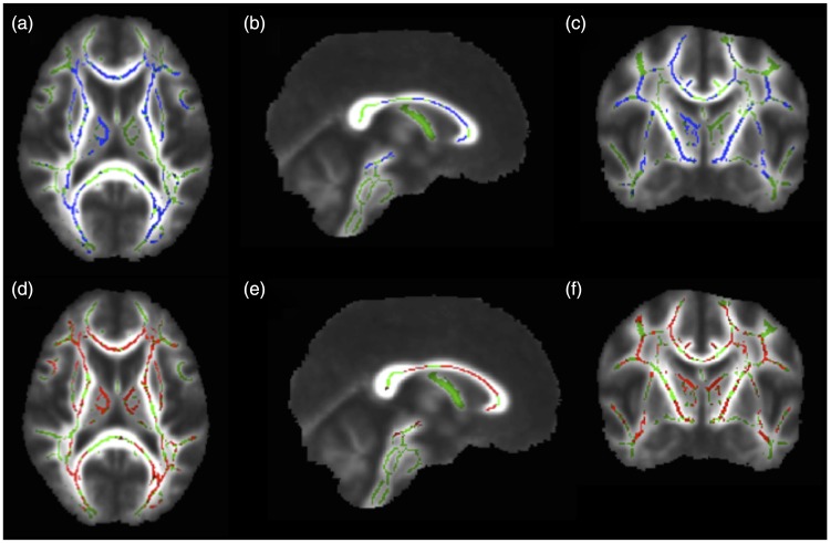
Figure 1.
Corrected probability maps showing voxels with significantly lower fractional anisotropy (FA) values in the brains of systemic lupus erythematosus (SLE) patients with memory deficit, compared to control individuals in blue (p < 0.05), in the axial (a), sagittal (b), and coronal (c) planes. Higher radial diffusivity (RD) values in the brain of SLE patients with memory deficit, compared to controls, are shown in red (p < 0.05) in the axial (d), sagittal (e), and coronal (f) planes. Note the large overlap of decreased FA and increased RD values in similar voxels of the brains of SLE patients with memory deficit, including bilateral anterior thalamic radiations, inferior fronto-occipital fasciculus, superior longitudinal fasciculus, uncinate fasciculus, corticospinal tract and corpus callosum.

