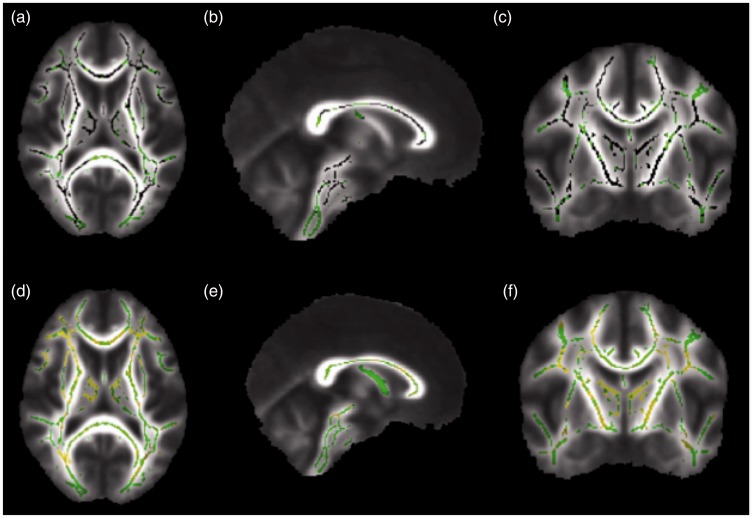
Figure 2.
Corrected probability maps showing voxels with significantly lower fractional anisotropy (FA) values in the brains of systemic lupus erythematosus (SLE) patients without memory deficit, compared to control individuals in black (p < 0.05), in the axial (a), sagittal (b), and coronal (c) planes. Higher radial diffusivity (RD) values in the brains of SLE patients without memory deficit, compared to controls, are shown in yellow (p < 0.05) in the axial (d), sagittal (e), and coronal (f) planes. There was substantial overlap of decreased FA and increased RD values in the brains of SLE patients without memory deficit, compared to controls. Compared to Figure 1, the diffusivity changes in the brains of SLE patients without memory deficit occurred in similar areas of the diffusivity changes in SLE patients with memory deficit, compared to the control group.

