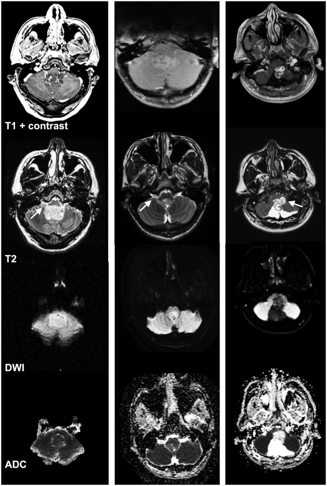
Figure 3.
Application of DWI and ADC histogram analysis for differentiation of fourth-ventricular tumors. Examples of three different fourth-ventricular tumors extending into the foramen of Luschka (arrows). The left column depicts a medulloblastoma with a 10th percentile ADC value of 568 × 10−6 mm2/s, the central column displays an ependymoma with a 10th percentile ADC value of 874 × 10−6 mm2/s, and the right column shows a pilocytic astrocytoma with a 10th percentile ADC value of 1487 × 10−6 mm2/s. The rows from top to bottom demonstrate postcontrast T1, T2, DWI, and ADC sequences. ADC: apparent diffusion coefficient; DWI: diffusion-weighted imaging.

