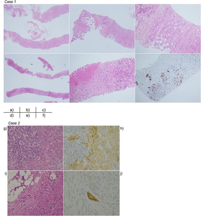
Fig. 2.
Pathological findings in Case 1 or Case 2. Case 1: (a) Breast biopsy specimen, hematoxylin-eosin (HE) stain ×20. (b) Breast biopsy specimen, HE stain ×40. (c) Breast biopsy specimen, HE stain ×100. (d) Liver necropsy specimen, HE stain ×20. (e) Liver necropsy specimen, HE stain ×100. (f) Liver necropsy specimen, gross cystic disease fluid protein 15 ×100. Case 2: (g) Liver biopsy specimen, hematoxylin-eosin (HE) stain ×20. (h) Liver biopsy specimen, E-cadherin stain ×40. (i) Breast biopsy specimen, HE stain ×10. (j) Breast biopsy specimen, E-cadherin stain ×40.

