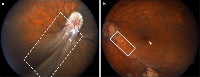
Fig. 1.
Fundus photographs of the posterior retina of the proband demonstrating FEVR. a Fundus picture of the right eye, showing a prominent falciform retinal fold (indicated with a dashed box) with several retinal vessels (indicated with white arrowheads) extending from the optic disk in inferior-temporal direction to the fundus periphery. The posterior retina is included in the fold and no macula could be detected. The retina outside the fold is attached and shows some retinal vessels. b Fundus picture of the left eye reveals areas of retinal pigmented epithelium (RPE) atrophy (indicated with a solid box) around the optic disk and along the inferior-temporal vessels. The macula (indicated with a white arrowhead) is slightly ectopic

