Fig. 2.
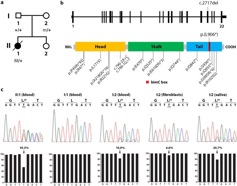
Genetic analysis of KIF11 in a patient with FEVR and microcephaly. a Pedigree of the family described in this study. The affected proband is depicted with a filled symbol. Slash indicates deceased person. M denotes germ line variant, m denotes mosaic variant. b Schematic representation of the KIF11 gene and the encoded Eg5 protein (adapted from Ostergaard et al. [7]). The variant identified in this study is depicted above the scheme. Other KIF11 variants reported in subjects with FEVR and microcephaly cases are indicated below the protein [4–6, 9]. cDNA positions corresponding to the indicated protein variants are listed in Table 1. c Sequencing results of the KIF11 variant. Sanger sequencing electropherograms are shown in the upper panel, whereas the relative amount of reads obtained from ion semiconductor sequencing are depicted below in bar graphs. The percentage of reads per given nucleotide is defined as the amount of reads called for a base among the total reads that cover that specific nucleotide. The percentage of reads having the c.2717del variant are indicated above the graph

