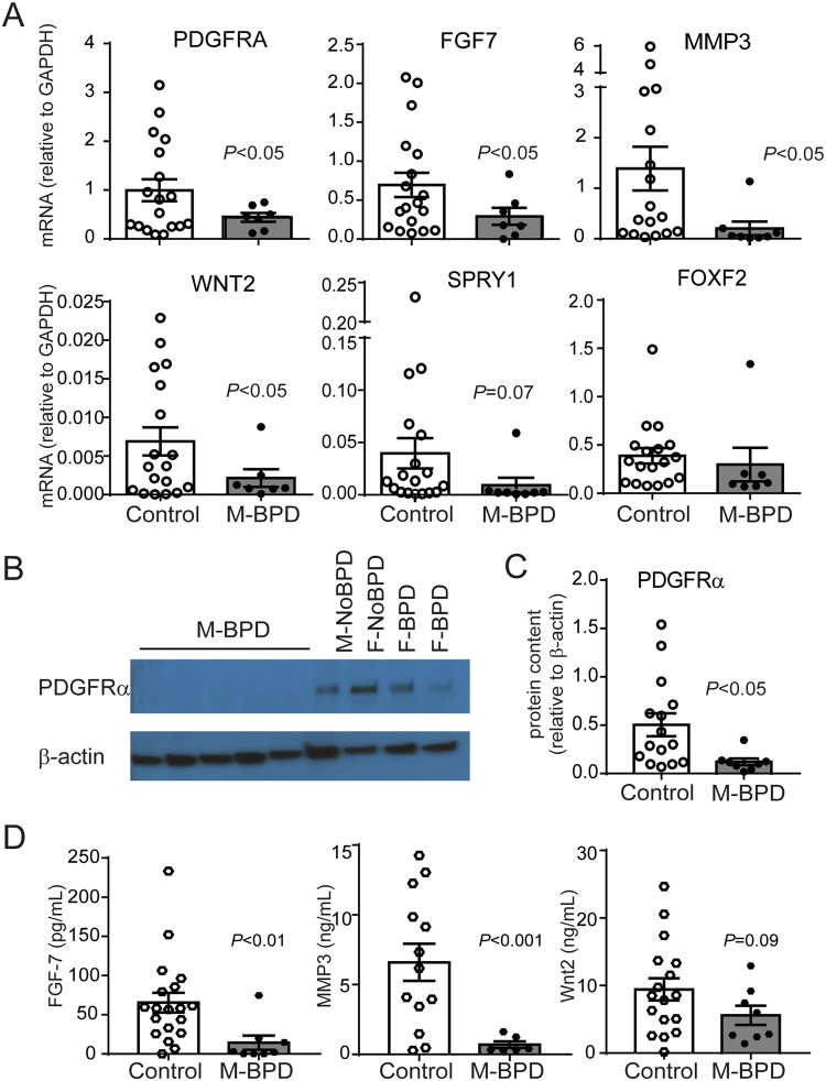
Figure 2.
Validation of the differential gene expression between MSCs from male infants developing BPD and combined controls. mRNA expression was assessed by quantitative PCR and protein expression was assessed by immunoblotting and ELISA. (A) Compared to the control group MSCs from male infants developing BPD showed significantly lower expression of PDGFRA, FGF7, WNT2 and MMP3 and a trend for lower SPRY1 and FOXF2 mRNA expression. (B) Representative immunoblot confirms decreased protein expression of PDGFRα. MSCs from 5 male infants developing BPD and 4 control infants (one male and one female infants who were not developing BPD and two female infants developing BPD) are shown. Full-length blots are available in Supplemental Fig. S1. (C) PDGFRα densitometry analysis group mean data for 9 male infants developing BPD and 18 controls. (D) MSCs from male infants developing BPD secrete significantly lower concentrations of FGF-7, MMP3 and Wnt2. Data are means ± SE. Statistical significance was determined by unpaired t-test.

