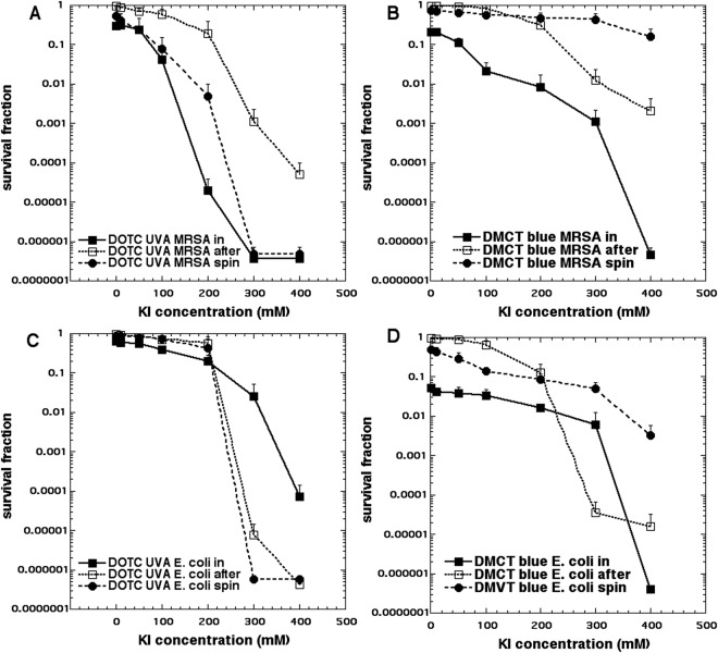
Figure 2.
Potentiation of aPDI by addition of KI. Bacteria (10(8) CFU/mL), tetracyclines (3 µM), exposed to 10 J/cm2 of UVA or blue light with the addition of different concentrations of KI. Cells were either present during light (in format), centrifuged before addition of KI and light (spin format), or added after light (after format). Controls (light alone or light + KI) showed no killing (data not shown). (A) Gram-positive MRSA with DOTC excited by UVA; (B) MRSA with DMCT excited by blue light; (C) Gram-negative E. coli with DOTC excited by UVA light; (D) E. coli with DMCT excited by blue light.

