. 2018 Nov 5;10(44):5273–5281. doi: 10.1039/c8ay01807c

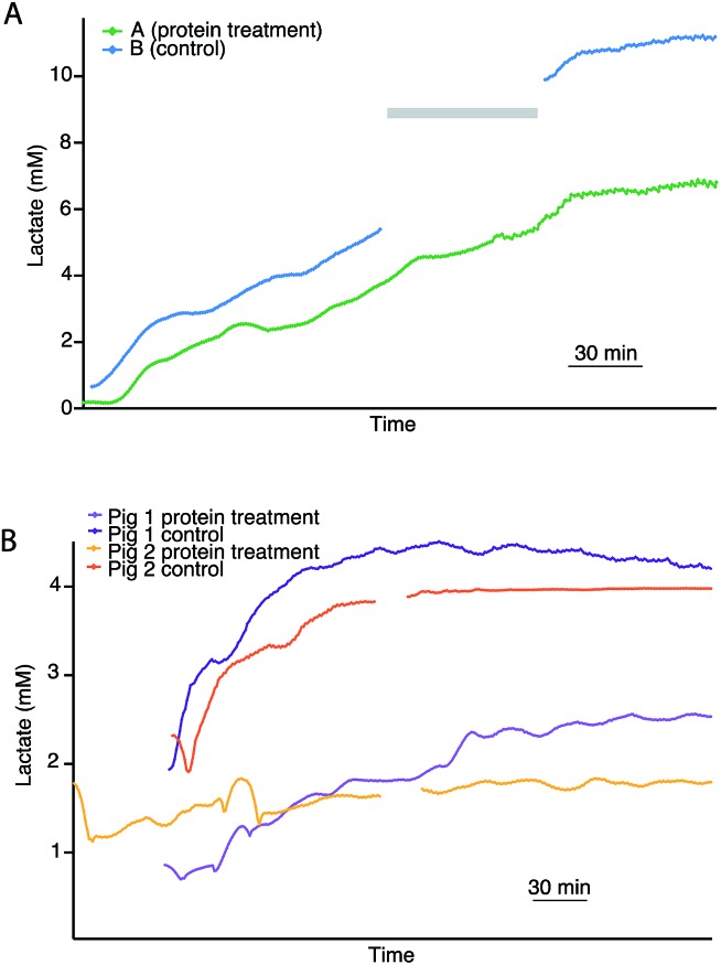
Fig. 4. (A) Dialysate cortical lactate profiles during haemoperfusion in paired human kidneys with (A) and without (B) thrombalexin pre-treatment. Both kidneys were previously subjected to 4 hours of HMP, after which kidney A (green) was treated with the novel cytotopic anticoagulant peptide thrombalexin and kidney B (blue) acted as the control. Following protein treatment, both kidneys were perfused at body temperature. Due to technical issues lactate levels were not recorded for kidney B during the time period indicated by a grey bar. (B) Dialysate cortical lactate profiles of two pairs of porcine kidneys undergoing haemoperfusion with and without protein pre-treatment. The plateau in lactate after 3.5 h haemoperfusion in the orange trace is caused by assay gain saturation at higher lactate levels. Each line represents real-time data of an individual kidney with a point every minute, smoothed with a Savitsky–Golay 21-point filter.

Fig. 4