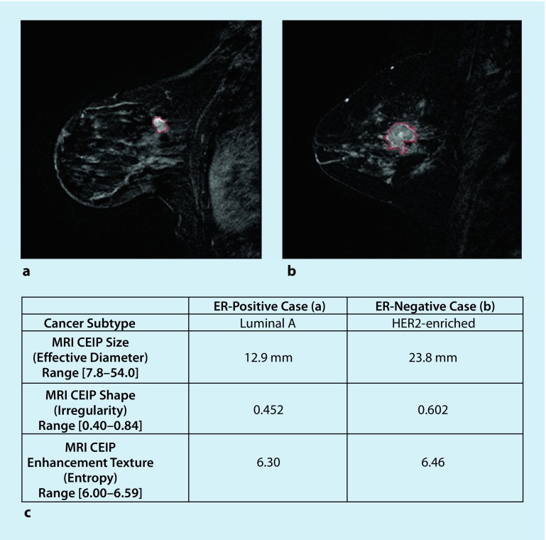
Fig. 2.
Example cases including segmentation outlines obtained from the computer segmentation method. a ER-positive example; b ER-negative example; c CEIP values (and ranges) for size, irregularity, and enhancement texture of two example cases. CEIP computer-extracted image phenotypes, ER estrogen receptor, HER2 human epidermal growth factor receptor 2, MRI magnetic resonance imaging. (Reprinted with no modifications under a creative common license (https://creativecommons.org/licenses/by/4.0/) from [29])

