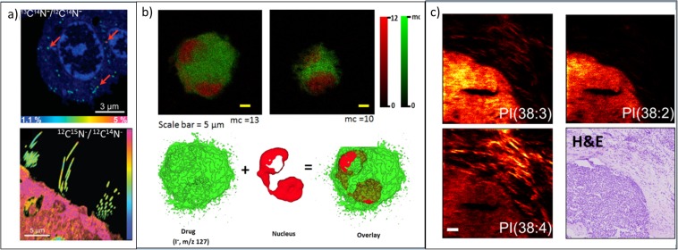
Fig. 4.
(a, upper) NanoSIMS image of the 13C14N−/12C14N− ratio image reveals the dopamine enrichment in single vesicles in pheochromocytoma (PC12) cells. Red arrows highlight three examples of vesicles. Reproduced from [34]. (a, lower) NanoSIMS image of 12C15N−/12C14N− ratio showing low incorporation of new proteins in stereocilia after feeding mice for 56 days with food containing 15N-leucine. Reproduced from [35]. (b, upper) ToF-SIMS images of NR8383 macrophage doped with desethylamiodarone (DEA) in negative ion. Snapshot images acquired using the Bi3+ ion beam for analysis and a GCIB for sample etching at different stages of the 3D image acquisition iodine, I− (green), and the summed ion contribution of the nuclear-markers (red). (b, lower) 3D isosurface rendering of the doped cell. I− is mapped in green and nuclear marker, HP2O6−, is mapped in red. Reproduced from [36]. (c) ToF-SIMS images of a human breast cancer biopsy using a (CO2)6k+ GCIB with H&E-stained image from a consecutive tissue slice. Three different phosphatidylinositol (PI) lipids distribute differently between the tumour and the inflammatory cells in the surrounding stroma. Reproduced from [37].

