Fig. 4.
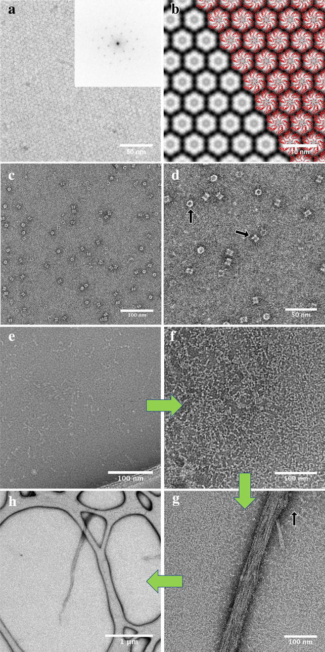
The applications of cell-free produced proteins in conventional electron microscopy. a STEM image of negatively stained 2D crystals of CCMK protein (inverted contrast). b The image from (a) has been processed using a temperature factor of − 100, and an IQ cutoff of 2 for the final map. The crystal exhibits P6 symmetry and a unit cell of a = b = 57 Å and γ = 120°. c, d TEM images of negatively stained PDX1.2 protein complexes. Top- and side-view orientations of the complex are depicted by black arrows. e–h TEM images of negatively stained SEO1 proteins. The individual fibril unit is indicated by a black arrow

