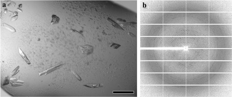
Fig. 6.
X-ray applications of cell-free-synthesized protein GS. a Optical image of 3D microcrystals for GS. The scale bar corresponds to 50 μm. b X-ray diffraction pattern collected from the crystals seen in (a). Data was collected at APS-NECAT beamline 24-ID-E with an EIGER 16 M pixel detector. The detector distance was 500 mm, and the wavelength was 0.9792 Å

