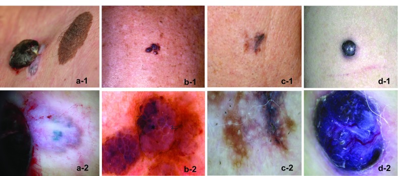
Fig. 2.

Images of four unusual subtypes of malignant melanoma. a Polypoid melanoma (Cabrera R, personal images). a-1 Close-up photograph of a 3-cm diameter, pedunculated pigmented lesion on the posterior trunk, in which the upper part is attached to the brown-blue pigmented skin with a colorless peduncle of only 0.5 cm. a-2 Dermatoscopy shows a blue-white veil in the exophytic lesion, and big blue-gray nests and whitish areas in the peduncle’s base. b Primary dermal melanoma (from Cabrera et al. [24]). b-1 Close-up photograph of a pigmented vascular lesion on the thorax. b-2 Dermatoscopy shows dark brown blotchy areas in some papillomatous projections (single arrow), areas with a light brown-pink color (double arrow), and a ‘blue-hue’ color without a veil (asterisk). c Verrucous malignant melanoma (Cabrera R, personal images). c-1 Close-up photograph of a verrucous lesion on the face. c-2 Dermatoscopy shows bluish-white veils with dots and globules, and peppering in some areas. d Pigmented epithelioid melanocytoma (from Aviles-Izquierda et al. [30]). d-1 Close-up photograph of a pigmented nodule on the arm. d-2 Dermatoscopy shows a homogeneous blue lesion with whitish structures, a blue-white veil, and polymorphic vessels
