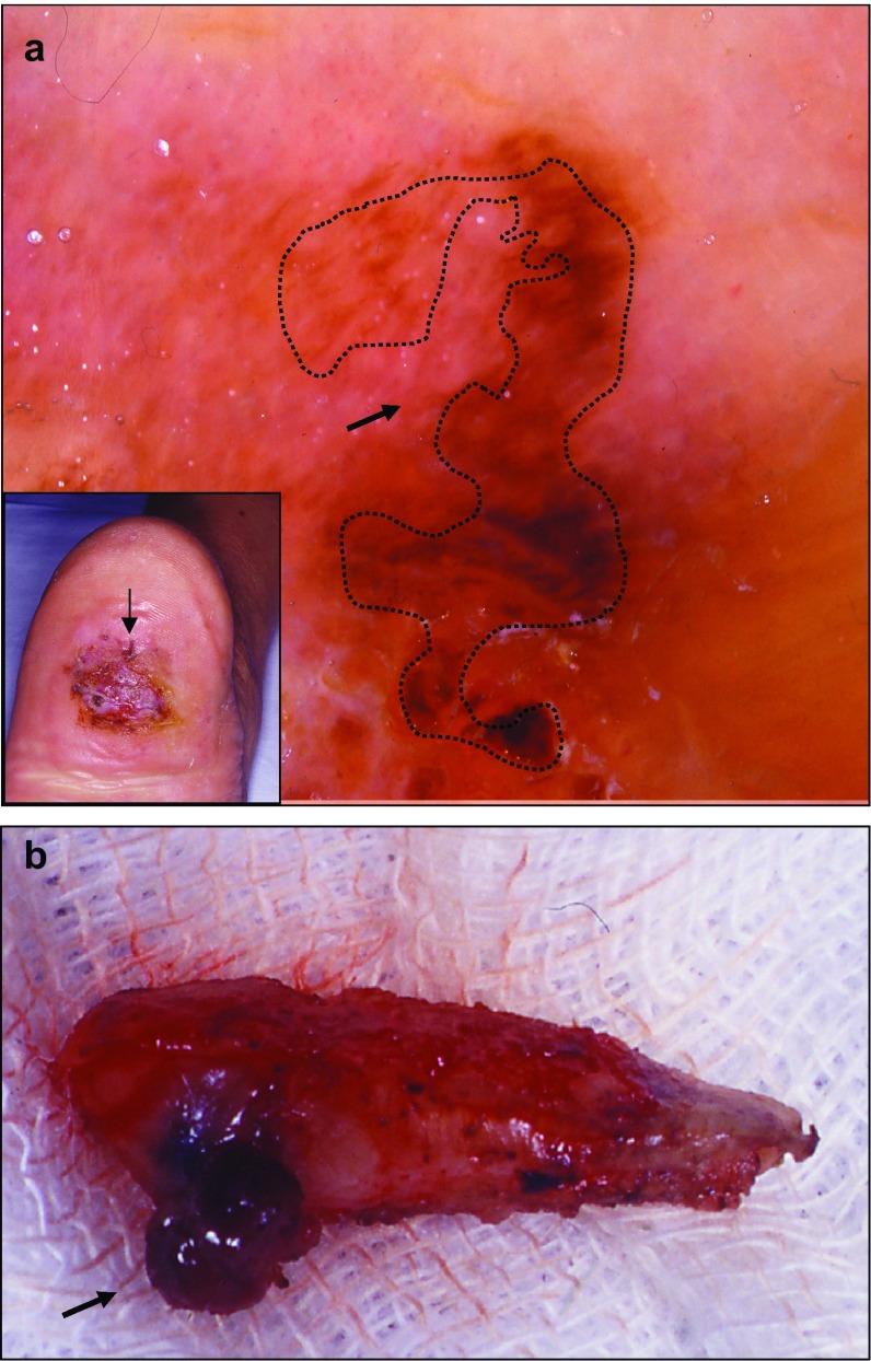
Fig. 5.
Hypomelanotic melanoma (Cabrera R, personal images). a Chronic foot ulcer with a hypopigmented lesion. In the inset, the arrow shows the exclusively peripheral localization of the atypical pigmented area on a chronic foot ulcer. Dermatoscopy of this area shows an atypical red pigmentation on the periphery (arrow on the main image). b Biopsy of the lesion. The deep part of the dermis presents pigmentation (arrow)

