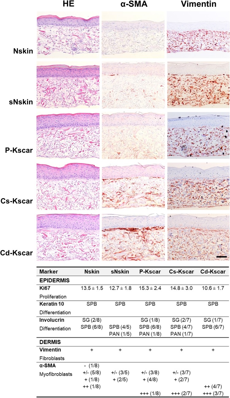
Fig. 3.
Increased α-SMA staining in central keloid regions. Upper panel shows representative pictures of HE, α-SMA and vimentin stainings performed on one of the duplicate skin models in n = 8 normal skin (Nskin), n = 8 peripheral keloid (P-Kscar), n = 7 central superficial keloid (Cs-Kscar), n = 7 central deep keloid (Cd-Kscar), n = 5 surrounding normal skin (sNskin). Magnification 200×, scale bar 100 µm. Lower panel shows the results of immunohistochemical stainings of epidermal markers (Ki67, keratin 10), dermal cellular markers (vimentin, α-SMA) in the skin models. Ki67 is expressed as mean ± SEM; SPB: suprabasal expression; SB: stratum basale; PAN: panepidermal (both SB and SPB); +/−: minimal expression; +: normal expression; ++: increased expression; +++: strongly increased expression; –: absent

