Figure 4.

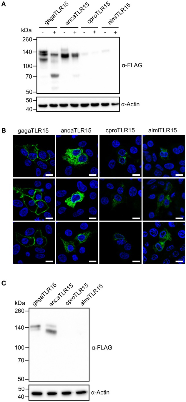
Proteolytic cleavage and expression of reptilian TLR15. (A) Immunoblot analysis of HEK293 cells expressing C-terminally FLAG-tagged chicken (gaga), anolis (anca), crocodile (cpro), or alligator (almi) TLR15 left untreated (–) or stimulated (+) (1 h) with 250 ng/mL Proteinase K. Mature TLR15 is ~140 kDa. Treatment with Proteinase K results in cleavage of gagaTLR15 and ancaTLR15 to form a cleaved receptor fragment that is slightly higher than 70 kDa. Note that crpoTLR15 and almiTLR15 are poorly expressed compared to ancaTLR15 and gagaTLR15. Beta-actin was detected to confirm equal loading of total protein onto SDS-PAGE gel. (B) Confocal microscopy on HEK293 cells expressing C-terminally HA-tagged TLR15 (green). Note that crpoTLR15 and almiTLR15 show lower expression compared to ancaTLR15 and gagaTLR15. All images were produced with the same microscopy settings. Nuclei are stained with DAPI (blue). White scale bar is 10 μm. Three representative images from two independent experiments are shown for each transfected group. (C) Immunoblot analysis of reptilian viper heart (VH-2) cells transfected with the different FLAG-tagged TLR15s. The rabbit α-human Beta actin antibody cross reacts with a specific protein in VH-2 cell lysate which was used to confirm equal loading of total protein onto SDS-PAGE gel. For (A,C); results are representative of three independent experiments.
