
Figure 3.
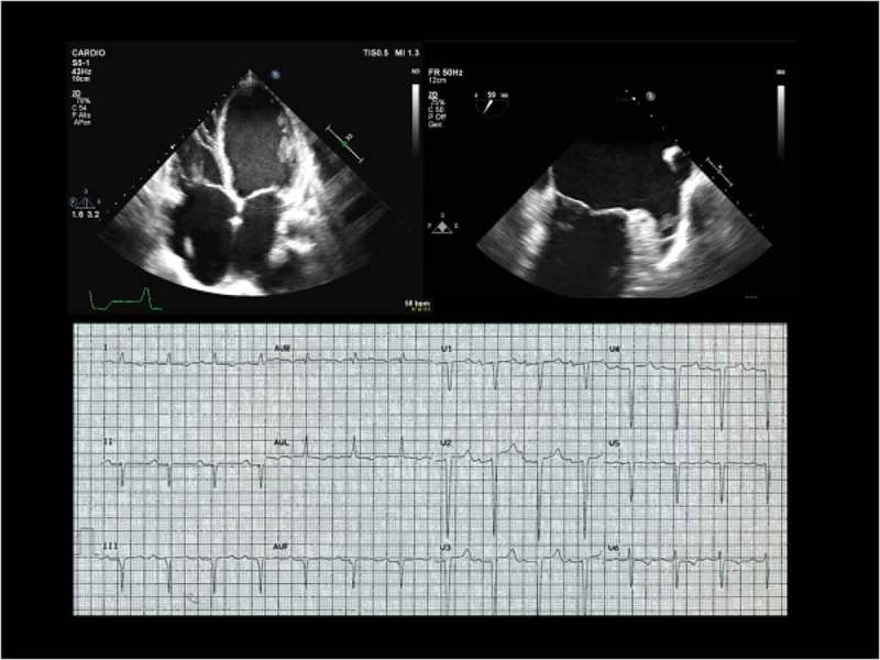
Echocardiographic (top) and electrocardiographic (bottom) findings of a typical cardiolaminopathy with paroxysmal atrial fibrillation. Panel A. Four-chamber view transthoracic echocardiography showing severe dilatation of both atria and severe dilatation and hypokinesia of both ventricles (ejection fraction of both left and right ventricles 20%). Panel B. Transesophageal echocardiography 60 degree view showing a trombus inside the left atrial appendage. Panel C. Electocardiogram: sinus rhythm, atrio-ventricular first degree A-V block, left anterior hemiblock and ventricular repolarization abnormalities.