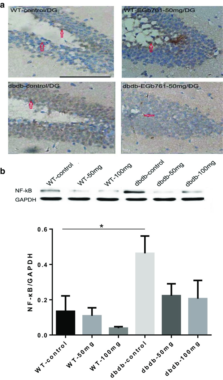
Fig. 6.
NF-κB immunohistochemistry staining. a Representative immunohistochemistry images of NF-κB staining. EGb761 reduced the expression of NF-κB protein in db/db−/− mice, but did not change NF-κB levels in WT mice. b The levels of NF-κB in the hippocampus were measured by western blotting. The graphs showed the relative levels of NF-κB to GAPDH. NF-κB expression was increased significantly in db/db−/− control compared with WT control (*, P < 0.05, F = 9.088). EGb761 reduced NF-κB levels in the db/db−/− groups without difference. n = 8 for WT 100 mg; n = 7 for WT 50 mg, db/db−/− control, and db/db−/− 100 mg; n = 5 for WT control and db/db−/− 50 mg

