Fig. 10.
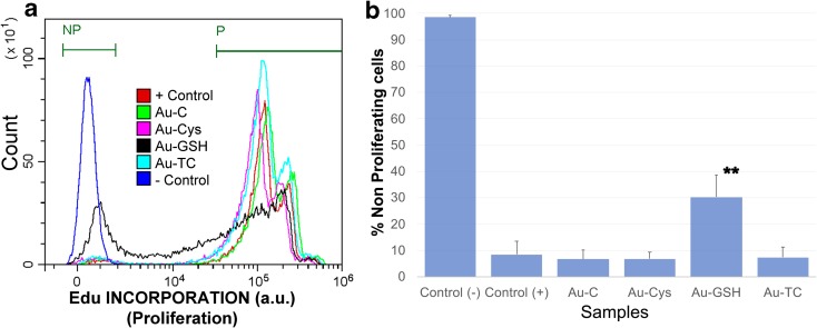
Detection of proliferating cells by EdU incorporation and labeling. Jurkat tumor cells were cultured with 10 μM EdU, in the presence or absence of the indicated nanoparticles at 15 μg/mL. Proliferation was determined as a function of Edu incorporation into cells’ DNA, detected by means of a Pacific blue fluorescent dye. EdU+ proliferating (gated in P) and non-proliferating cells (NP) were clearly and distinctly separated by FACS. Jurkat cells in the absence of NP were used as a positive control (+ control) for proliferation, as they are a tumor cell line that spontaneously proliferate in culture. Cells that have not incorporated Edu were used as negative control (− control). a Overlay histogram showing the proliferation profile of all samples. Samples had been electronically gated according to their size/granularity distribution. Individual samples as well as the gating strategy are shown in supplementary Fig. S.3. An overlay histogram of the ungated populations as well as results corresponding to individual ungated samples is shown in Fig. S.4.b) Average and standard errors for the percentage of non-proliferating cells under each condition (**p < 0.001)

