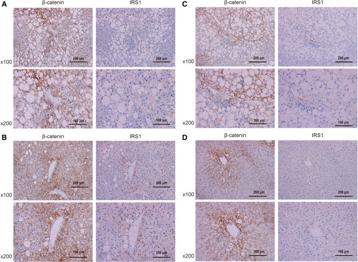
Fig. 5.
Immunohistochemical staining of liver biopsies from representative nonalcoholic fatty liver disease patients. Patients A and B underwent liver biopsy 5 h after oral glucose tolerance tests (OGTT), whereas patients C and D underwent liver biopsy in a fasting state. a Patient A was a 44-year-old male with severe ballooning. b Patient B was a 31-year-old male, with no ballooning. c Patient C was a 47-year-old male and had severe ballooning. d Patient D was a 45-year-old male and had no ballooning. Liver biopsies were stained for β-catenin and IRS1. Positive immunoreactivity appears brown. Original magnification, ×100 or ×200

