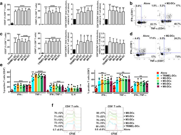
Fig. 4.
Activation of allogeneic T cells by monocyte-derived DCs matured with different heat shock-conditioned GBC lysates. Purified CD3+ T cells were co-cultured for 5 days with allogeneic TRIMEL-, M2-, M3-, M5-, M8-DCs or without DCs. The surface expression of CD25, CD69, CXCR3 and CXCR4 (a), the intracellular levels of IFN-γ, TNF-α and IL-4 (b–d), and proliferation (e) were evaluated in the CD4+ and CD8+ T cells populations by flow cytometry. a, d Bars represent the average and SD from five independent experiments of the % of T cells positive for each marker, with the exception of CXCR3 and CXCR4 data that are shown as fold induction of the MFI relative to unstimulated T cells. Representative dot plots of IFN-γ and TNF-α production in allogeneic CD4+ (b) and CD8+ (c) T cells co-cultured with M2-DCs. e The percentage and SD of proliferating T cells are showed on the left of each histograms. Evaluated cell lysates mix were made as follows: M2 (2TKB + 24TKB + GBd1); M3 (1TKB + 2TKB + 24TKB); M5 (2TKB + G415 + OCUG1); and M8 (24TKB + OCUG1 + G415). *p < 0.05; **p < 0.01; ***p < 0.001; ****p < 0.0001 (comparison versus unstimulated T cells)

