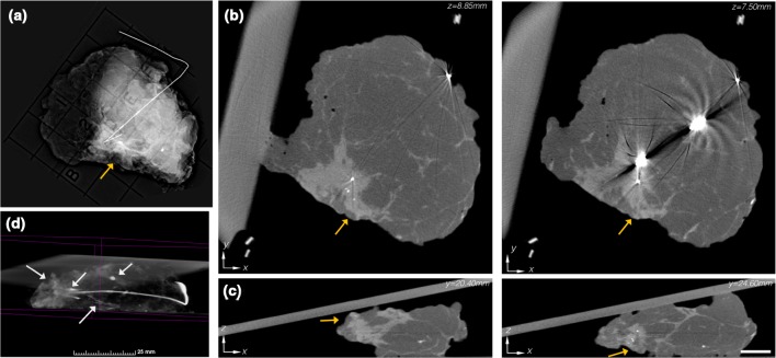
Fig. 2.
a Single-projection specimen mammogram revealing a peripherally located IDC + DCIS lesion (orange arrow). b Reconstructed micro-CT slices (1.35 mm apart) similarly depict the peripherally located mass. Notable metal reconstruction artifacts occur because of the localization wire. c Orthogonal micro-CT slices (4.2 mm apart) suggest a close peripheral margin, and also indicate a close deep margin, which would not be evident in the single-projection specimen mammogram. Micro-CT scale bar is 1 cm. d 3D volume rendering of the specimen, with the adipose tissue thresholded to be transparent, revealed a micro-calcification cluster, a large smooth calcification, tumor location metal clip, and calcified vasculature (white arrows) in the presentation (see video in Additional File 3)

