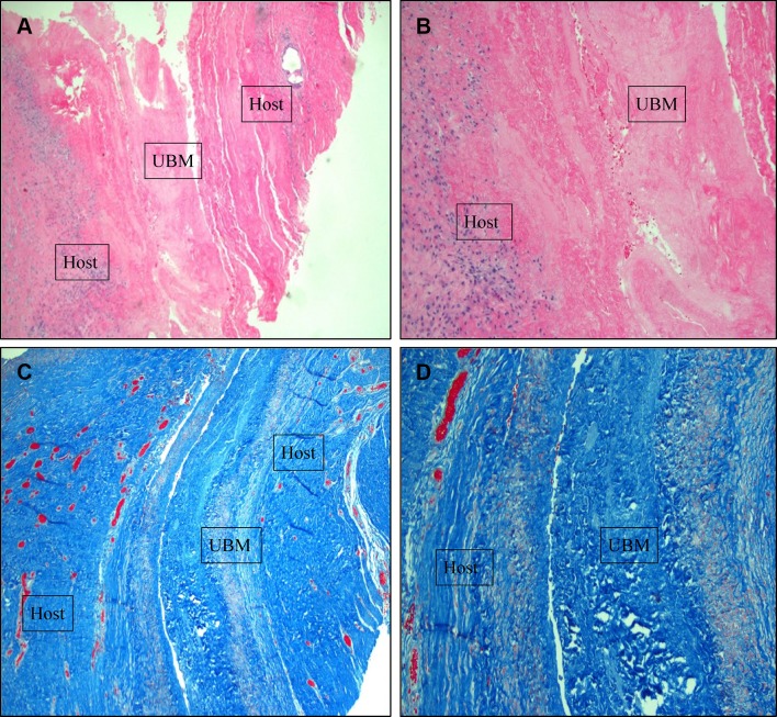
Fig. 5.
Myofascial biopsies 14 months after retrorectus repair of complex incisional hernia with UBM reinforcement. a 4× power H and E staining showing native and graft tissues approximated, b 10× power H&E staining showing remodeling response, c 4× power Trichrome stain demonstrating the retrorectus position of the xenograft, and d 10× power Trichrome stain demonstrating the remodeling response at the interface between the host and graft layers

