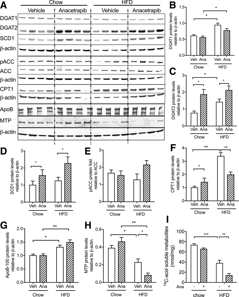
Figure 6.
CETP inhibition promoted TG synthesis and impaired fatty acid oxidation in the liver in HFD-fed CETP mice. A: Western blots of liver DGAT1, DGAT2, SCD1, pACC, ACC, CPT1, apoB, and MTP from chow- and HFD-fed mice. β-actin was used as the loading control. B–D, F–H: Quantification of Western blots for each protein. E: Ratio of pACC to ACC protein amounts in the liver. I: Fatty acid oxidation was measured with primary hepatocytes from CETP male mice that were treated with anacetrapib or vehicle. Data are mean ± SD (n ≥ 6; n = 4 in I). Significant differences were determined using repeated-measures by both factors two-way ANOVA with Bonferroni multiple comparison test. *P < 0.05, **P < 0.01, ***P < 0.001. Ana, anacetrapib; Veh, vehicle.

